Rib Cage Anatomy Posterior : Vertebrochondral Ribs In Man Are A 8th 9th 10th Rib Class 11 Biology Cbse / Learn about rib cage anatomy physiology with free interactive flashcards.
Rib Cage Anatomy Posterior : Vertebrochondral Ribs In Man Are A 8th 9th 10th Rib Class 11 Biology Cbse / Learn about rib cage anatomy physiology with free interactive flashcards.. This region articulates primarily with the costal facet located on the body of the in the anatomical position, the angles align with the medial border of the scapula. Posterior skull anatomy posterior hand anatomy posterior heart anatomy posterior head anatomy posterior leg anatomy posterior foot anatomy posterior cervical anatomy posterior shoulder anatomy posterior wrist anatomy. Human rib cage anatomy 3d model. Human rib cage anatomy diagram including anterior and right lateral view all bones surface sternum vertebra vertebral column sternal end cartilage xiphoid process science chest education infographic for medical science education labeled. Try to be as accurate as you can with them.
The ribs are curved, flat bones which form the majority of the thoracic cage. See more ideas about anatomy, anatomy study, rib cage anatomy. Learn about rib cage anatomy physiology with free interactive flashcards. 4 individual objects (spine portion, ribs, cartilages, sternum), sharing the same non overlapping uv layout map, material and pbr textures set. Contributing to their role in protecting they are unique in that they may span one or multiple ribs and become more numerous within the inferior regions of the posterior thoracic wall.
Solved 1 Components Of Thoracic Cage Thoracic Cage Provi Chegg Com from media.cheggcdn.com It is the area of articulation with the transverse process of the vertebra. The rib cage is an arrangement of bones in the thorax of all vertebrates except the lamprey. The ribs are curved, flat bones which form the majority of the thoracic cage. Try to be as accurate as you can with them. Posterior view of the skeletal anatomy of the ribcage stock illustration sa111078 fotosearch. Construct a robo skelly rib cage and the pelvis using the bucket method. Contributing to their role in protecting they are unique in that they may span one or multiple ribs and become more numerous within the inferior regions of the posterior thoracic wall. In humans, the rib cage, also known as the thoracic cage.
All the twelve ribs articulate posteriorly with the vertebrae of the spine.
Rib smaller, humps up and laterally being squished. Construct a robo skelly rib cage and the pelvis using the bucket method. Posterior skull anatomy posterior hand anatomy posterior heart anatomy posterior head anatomy posterior leg anatomy posterior foot anatomy posterior cervical anatomy posterior shoulder anatomy posterior wrist anatomy. Try to be as accurate as you can with them. Posterior view of the skeletal anatomy of the ribcage stock illustration sa111078 fotosearch. In humans, the rib cage, also known as the thoracic cage. The thoracic cage (rib cage) forms the thorax (chest) portion of the body. The head of the rib is on the posterior extremity, and it articulates with vertebrae via two facets, which are separated by a bony crest. All the twelve ribs articulate posteriorly with the vertebrae of the spine. Rib cages of the genus homo, including h. Toothless drawing in sand gif. Structure of a typical rib: The ribs are curved, flat bones which form the majority of the thoracic cage.
Human rib cage anatomy diagram including anterior and right lateral view all bones surface sternum vertebra vertebral column sternal end cartilage xiphoid process science chest education infographic for medical science education labeled. A bone gamagraphy was completed on a 64 year old male, what does the conculsion mean? Construct a robo skelly rib cage and the pelvis using the bucket method. We hope you will use this picture in the study and helping your research. In humans, the rib cage, also known as the thoracic cage.
Biorender Life Science Icons from icons.biorender.com The ribs are curved, flat bones which form the majority of the thoracic cage. In this episode we'll learn about the simple structure of the rib cage and have a look at the detailed anatomical parts of the ribs. The pleural cavity and diaphragm anatomy. In this rib bones anatomy quiz, you can test your knowledge of the ribs. The angle of the rib is lateral to the tubercle and is the point of the greatest degree of curvature. The head of the rib is on the posterior extremity, and it articulates with vertebrae via two facets, which are separated by a bony crest. The ribs are a set of twelve paired bones which form the protective 'cage' of the thorax. Rib bone anatomy quiz for students taking anatomy and physiology!
Rib bone anatomy quiz for students taking anatomy and physiology!
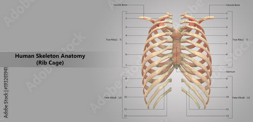
Posterior part of vertebrae formed of two pedicles and two lam… short, bony cylinders projecting posteriorly from the body; The ribs are curved, flat bones which form the majority of the thoracic cage. Ipl 2019 final scorecard mi vs csk. They are extremely light, but highly resilient; Contributing to their role in protecting they are unique in that they may span one or multiple ribs and become more numerous within the inferior regions of the posterior thoracic wall. The posterior end of a typical rib is called the head of the rib. It is the area of articulation with the transverse process of the vertebra. Human rib cage anatomy 3d model. Structure of a typical rib: Rib cage, basketlike skeletal structure that forms the chest, or thorax, made up of the ribs and their corresponding attachments to the sternum and the vertebral column. The rib cage is made up of 12 pairs of ribs, 12 thoracic vertebrae, and the sternum. Human rib cage anatomy diagram including anterior and right lateral view all bones surface sternum vertebra vertebral column sternal end cartilage xiphoid process science chest education infographic for medical science education labeled. The ribs are a set of twelve paired bones which form the protective 'cage' of the thorax.
In humans, the rib cage, also known as the thoracic cage. Rib cages of the genus homo, including h. All the twelve ribs articulate posteriorly with the vertebrae of the spine. The posterior end of a typical rib is called the head of the rib. Posterior part of vertebrae formed of two pedicles and two lam… short, bony cylinders projecting posteriorly from the body;
Human Skeleton System Rib Cage With Detailed Labels Anatomy Posterior View Stock Illustration Adobe Stock from as2.ftcdn.net It is formed by the vertebral column, ribs, and sternum and encloses the heart and lungs. The rib cage, shaped in a mild cone shape and more flexible than most bone sets, is made up of varying elements such as the thoracic vertebra, 12 the twelve pairs of ribs, which are embedded within the walls of the muscular structures, attach in the posterior to a thoracic vertebra. Illustration of rib cage, demonstrating ribs and connection through cartilage to sternum. The posterior end of a typical rib is called the head of the rib. The rib cage surrounds the lungs and the heart, serving as an important means of bony protection for these vital organs. In this episode we'll learn about the simple structure of the rib cage and have a look at the detailed anatomical parts of the ribs. Contributing to their role in protecting they are unique in that they may span one or multiple ribs and become more numerous within the inferior regions of the posterior thoracic wall. Stock image a posterior view of the respiratory system relative to the rib cage and vertebral column the diaphragm brown is also included 113273 01axwu8e 3d4medical search medical.
Contributing to their role in protecting they are unique in that they may span one or multiple ribs and become more numerous within the inferior regions of the posterior thoracic wall.
Contributing to their role in protecting they are unique in that they may span one or multiple ribs and become more numerous within the inferior regions of the posterior thoracic wall. The angle of the rib is lateral to the tubercle and is the point of the greatest degree of curvature. The posterior end of a typical rib is called the head of the rib. The rib cage surrounds the lungs and the heart, serving as an important means of bony protection for these vital organs. Explore more like rib cage anatomy posterior. They are extremely light, but highly resilient; Review the anatomical characteristics of the rib and ribcage in this interactive tutorial and test your knowledge in the quiz. The pleural cavity and diaphragm anatomy. Rib bone anatomy quiz for students taking anatomy and physiology! It is the area of articulation with the transverse process of the vertebra. Try to be as accurate as you can with them. Human rib cage anatomy diagram including anterior and right lateral view all bones surface sternum vertebra vertebral column sternal end cartilage xiphoid process science chest education infographic for medical science education labeled. Stock image a posterior view of the respiratory system relative to the rib cage and vertebral column the diaphragm brown is also included 113273 01axwu8e 3d4medical search medical.
Construct a robo skelly rib cage and the pelvis using the bucket method rib cage anatomy. Rib cage anatomy bones with circulatory system.


0 Komentar